Home
Uncategories
Upper Leg Tendon Anatomy / Upper Leg Muscles Toxinmed / Your lower leg includes three main muscles, located behind your tibia or shinbone.
Upper Leg Tendon Anatomy / Upper Leg Muscles Toxinmed / Your lower leg includes three main muscles, located behind your tibia or shinbone.
Upper Leg Tendon Anatomy / Upper Leg Muscles Toxinmed / Your lower leg includes three main muscles, located behind your tibia or shinbone.. This mri wrist coronal cross sectional anatomy tool is absolutely free to use. Upper leg muscle pain is a very hard pain affect the leg pain as a whole. The thigh muscles don't just move your legs. And it is also critical to the walking process. Chest muscle anatomy exercises 12 photos of the chest muscle anatomy exercises chest muscle anatomy and exercises, chest muscle anatomy exercises, human muscles, chest muscle anatomy and exercises, chest muscle anatomy exercises
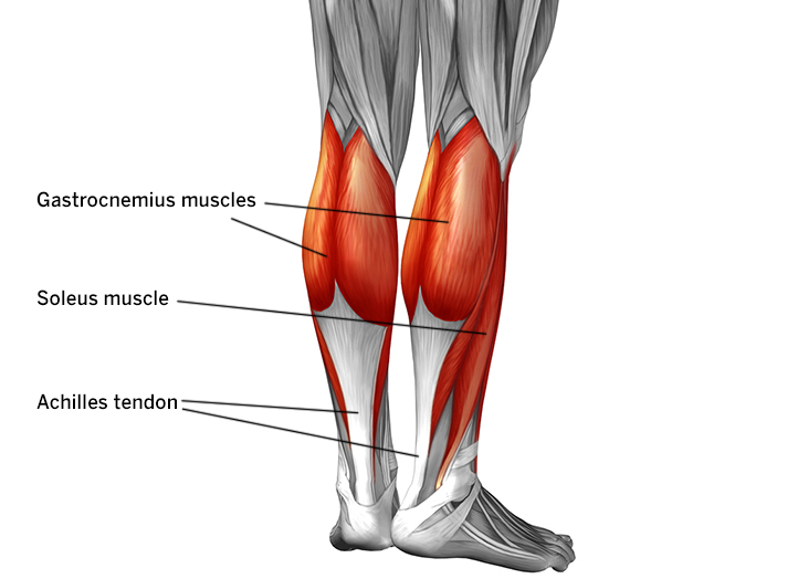
Upper leg tendon anatomy : The calf comprises of 2 major muscles (gastrocnemius and soleus) both of which. The achilles tendon or heel cord, also known as the calcaneal tendon, is a tendon at the back of the lower leg, and is the thickest in the human body. Meanwhile, the vastus lateralis is on the side of the thigh, while the vastus intermedius is hidden below the rectus femoris(5). Related posts of muscle anatomy upper leg.
Achilles Tendon Pain Causes Diagnosis And Treatment from www.hss.edu Upper leg muscle pain is a very hard pain affect the leg pain as a whole. And it is also critical to the walking process. On the medial edge of the posterior thigh is the gracilis muscle. Upper leg anatomy and function the upper leg is often called the thigh. This is why you have to indicate which biceps you are taking about when discussing one or other of these muscles. The rectus femoris is located in the center of the thigh, while the vastus medialis is in the middle of the said body part. They have a lot to do with how your hips move. The muscles of the thigh and gluteal region are a group of complex muscles that help move and stabilize the lower limb.
The iliopsoas is a workaholic muscle.
Anterior muscles extend your legs and flex your thighs. Other muscles of the anterior (front) thigh include the pectineus, sartorius,. Squeeze your knees together and boom, you're contracting the adductors. The human leg, in the general word sense, is the entire lower limb of the human body, including the foot, thigh and. Tendons are cords made of tough tissue, and they work as special connector pieces between bone and muscle. It is also visible on the medial edge of the thigh from the anterior. The thigh muscles don't just move your legs. The iliopsoas is a workaholic muscle. This is the group of muscles that you often see body builders flexing, which protrude just above the knee and take up most of the upper leg. Tendon, tissue that attaches a muscle to other body parts, usually bones. The achilles tendon or heel cord, also known as the calcaneal tendon, is a tendon at the back of the lower leg, and is the thickest in the human. The human leg, in the general word sense, is the entire lower limb of the human body, including the foot, thigh and even the hip or. Related posts of muscle anatomy upper leg.
Its muscle belly is on the back aspect of the upper arm. The muscles of the thigh and gluteal region are a group of complex muscles that help move and stabilize the lower limb. The human leg, in the general word sense, is the entire lower limb of the human body, including the foot, thigh and. The hamstrings refer to 3 long posterior leg muscles, the biceps femoris, semitendinosus, and semimembranosus. Tendons are cords made of tough tissue, and they work as special connector pieces between bone and muscle.
Muscle Anatomy from droualb.faculty.mjc.edu Standing radiographs are shown in figures a and b. This is why you have to indicate which biceps you are taking about when discussing one or other of these muscles. Related posts of muscle anatomy upper leg. The iliopsoas is a workaholic muscle. Upper leg tendon anatomy : Upper leg tendon anatomy : Lateral (fibular) collateral ligament (fcl) upper part middle part lower part popliteus tendon (pt) upper part i. Anterior muscles extend your legs and flex your thighs.
Its muscle belly is on the back aspect of the upper arm.
And it is also critical to the walking process. The knee joint is commonly injured, so understanding its anatomy can help you understand the conditions that cause problems, so you stay safe and prepared. It is also visible on the medial edge of the thigh from the anterior. Related posts of muscle anatomy upper leg. It also is active in maintaining thigh and kneecap position while walking and. It serves to attach the plantaris, gastrocnemius (calf) and soleus muscles to the calcaneus (heel) bone. Tendon, tissue that attaches a muscle to other body parts, usually bones. These muscles run from the lower spine and pelvis, join together, then attach by a tendon to the upper thigh. Upper leg tendon anatomy : The calf comprises of 2 major muscles (gastrocnemius and soleus) both of which. Medial muscles adduct and rotate your thigh, and posterior flex your leg and extend your thigh. This important tendon in the back of the calf and ankle connects the plantaris, gastrocnemius, and soleus muscles to. Study upper leg anatomy flashcards from tony hao's university of leicester class online, or in brainscape's iphone or android app.
Notice the upper leg has a biceps muscle just like the upper arm does. Pin on upper leg muscle anatomy from i.pinimg.com there is no real division between the core and the upper leg; Anatomy the four quadriceps muscles meet just above the kneecap (patella) to form the quadriceps tendon. For more on tendon anatomy, refer here. Related posts of muscle anatomy upper leg.
Upper Leg Muscles Toxinmed from www.toxinmed.com Pin on upper leg muscle anatomy from i.pinimg.com the human leg, in the general word sense, is the entire lower limb of the human body, including the foot, thigh and even the hip or gluteal region. This important tendon in the back of the calf and ankle connects the plantaris, gastrocnemius, and soleus muscles to. Standing radiographs are shown in figures a and b. Related posts of muscle anatomy upper leg. The achilles tendon or heel cord, also known as the calcaneal tendon, is a tendon at the back of the lower leg, and is the thickest in the human body. The achilles tendon or heel cord, also known as the calcaneal tendon, is a tendon at the back of the lower leg, and is the thickest in the human. Anterior muscles extend your legs and flex your thighs. The quadriceps tendon attaches the quadriceps muscles to the patella.
Squeeze your knees together and boom, you're contracting the adductors.
Your upper leg includes seven major muscles. In clinical anatomy the thigh muscles are divided into three groups: They consist of the rectus femoris, vastus intermedius, vastus lateralis and the vastus medialis. The achilles tendon or heel cord, also known as the calcaneal tendon, is a tendon at the back of the lower leg, and is the thickest in the human body. Its muscle belly is on the back aspect of the upper arm. The fibers run vertically downward, and end in a rounded tendon, which passes behind the medial condyle. Tendons are thick bands of tissue that connect muscles to bone. The achilles tendon or heel cord, also known as the calcaneal tendon, is a tendon at the back of the lower leg, and is the thickest in the human body. Upper leg muscles art print barewalls posters prints bwc13091068 / muscles of the leg 3d interactive anatomy tutorial originates from the common tendon and attaches to the upper spine and skull. This is why you have to indicate which biceps you are taking about when discussing one or other of these muscles. Tendons are cords made of tough tissue, and they work as special connector pieces between bone and muscle. The achilles tendon or heel cord, also known as the calcaneal tendon, is a tendon at the back of the lower leg, and is the. The four muscles all extend the lower leg.


0 Comments:
Post a Comment